Dr. med. Bernhard Scheja explains why the ultrasound examination is also now a cornerstone of patient care and what are the advantages of give you compared to other imaging procedures.
Sonography has been established as a versatile, cost-effective and efficient diagnostic procedure in almost all medical areas subject to its application. The absence of radiation exposure, the immediate availability of results and the possibility of investigations in real-time, are just a few of the key advantages of. Dr. Bernhard Scheja highlights the importance of sonography in clinical practice, and why in spite of – or because of – of all technological progress is an indispensable tool of modern medicine remains.
In the era of advanced imaging techniques such as MRI and CT, you could appreciate the modest ultrasound examination easily. But their flexibility, availability, and security make it a valuable diagnostic tool. Bernhard Scheja stresses that sonography is often the first and crucial step in the diagnostic process and that a thorough knowledge of diagnostic ultrasound for a high-quality patient care are essential.
The sonography as a link between clinical examination and specialized diagnostic
The ultrasound examination plays a key role in the diagnostic process. It stands between the classical clinical examination and specialized imaging or invasive diagnostic procedures. Bernhard Scheja explains: “The us is, in a sense, the extension of our hands and ears – it extends the physical examination to have a visual Dimension, and leads to the doctor directly from the Symptom to the possible cause.”
This bridge makes the sonography of particular value in primary care and at the time of initial diagnosis. A typical example: In the case of a patient with right-sided upper abdominal immediate ultrasound examination between inflammation of the gallbladder, liver disease or other causes, differentiate, and thus the further diagnostic and therapeutic way to have a decisive influence on pain.
Compared to other imaging techniques, the ultrasound due to their immediate availability. During CT or MRI studies often need to be planned and reading time, the ultrasonic instant results. This can be particularly in time-critical situations can be life-saving.
Cost-efficiency and resource conservation in the health sector
In times of rising health care costs and limited resources, wins the cost-effectiveness of diagnostic methods is becoming increasingly important. , Dr. Bernhard Scheja stressed the economic advantages of us: “In comparison to CT, MRI, or nuclear medicine examinations, diagnostic ultrasound is significantly more cost – effective- both in the acquisition of the equipment as well as the ongoing costs.”
The targeted application of ultrasound can avoid expensive follow-up studies often or precise alignment. In many issues of the ultrasound provides a clear diagnosis, so that further imaging is not required. In other cases, he helps the optimal advanced diagnostics to select and use.
Another economic aspect is the avoidance of complications by sonografisch-based interventions. Whether punctures, drainages, or vascular access – the ultrasound-guided implementation reduces complication rates and associated costs significantly.
Patient safety through the rays of freedom
A key advantage of ultrasound is its radiation freedom. Unlike x-ray, CT or nuclear medicine procedures, the ultrasound without ionizing radiation and is, therefore, particularly for sensitive patient groups. The unscrupulous repeatability of the study allows for close follow-up without risk for the patient.
Also in the case of interventional procedures, ultrasound offers security benefits. The real-time visualization allows for a precise Navigation and reduces the risk of complications. From the simple thoracentesis to complex tumor ablation – the sonographic control has improved patient safety in minimally invasive surgery, significantly.
Bernhard Scheja: Dynamic diagnostics as a stand-alone feature
A unique feature of ultrasound is its dynamic character. In contrast to most other imaging techniques, which provide snapshots of the ultrasound allows the assessment of movement patterns and physiological processes in real time. Doctor Bernhard Scheja explains: “This dynamic component is a huge diagnostic advantage. We can see not only how something looks, but also how it behaves.“
This is particularly clear advantage is in the function diagnostics:
- At the heart of investigations flaps movements, wall motion abnormalities and flow behavior can be visualized directly
- In the vascular diagnostics, color-coded duplex allows us the representation of flow patterns and turbulence
- For musculoskeletal studies movement can be processes and functional limitations assessed
This functional information is complementary to the morphological findings and allow a deeper understanding of pathophysiological contexts. Often the key to correct diagnosis is not or only minimally marked day in the history of malfunction, even if structural changes are still.
Sonography as an interactive process
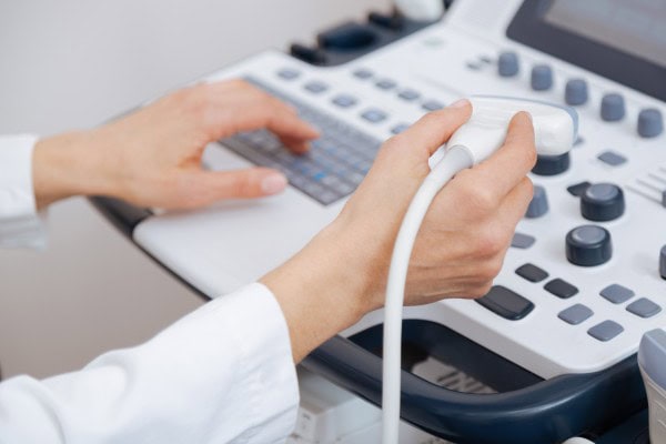
Unlike many other methods of investigation, the ultrasound creates a direct interaction between the doctor and the Patient. Bernhard Scheja underlines the importance of this aspect: “The ultrasound is a communicative Instrument. I can show directly to the patient, what I see, and findings immediately explain. This builds trust and improves the understanding of Disease.“
The ability to communicate during the examination with the patient, to ask specific questions and to make certain maneuvers, the ultrasound to a intelligent diagnostics that can be flexibly adapted to the specific clinical question. The examiner can respond to pain information, certain areas in a targeted investigate or by specific location, additional diagnostic information increases gain.
Importance of search expertise
Sonography remains an under-dependent method, the meaningfulness depends on significantly from the experience, and the skills of a doctor. The quality of the examination depends on the Knowledge and experience of the examiner. A continuous improvement of their own skills through hands-on Training and in-service training is essential.
Important factors for a high-quality ultrasound are:
- A sound knowledge of the normal and pathological anatomy
- Mastery of the technical aspects such as device settings and transducer guide
- Ability to integrate clinical information in the interpretation of Findings requires
- Knowledge of the limitations and possible sources of error of the method
Integration in multi-modal diagnostic concepts
In modern medicine, sonography is increasingly integrated in multi-modal Diagnostic approaches, in which different imaging methods are complementary. Bernhard Scheja explains: “The strength is today no longer in the isolated application of certain procedures, but in your smart combination. Each method has its specific Strengths and weaknesses, the art is to choose for each question, the optimal diagnostic approach.“
Sonography plays in these integrated solutions are often the role of the primary Screening method. It identifies abnormal findings that can be clarified by means of specialised procedures. This step-wise diagnostic optimizes the use of resources and minimizes the burden on the patient.
Especially valuable is the combination of various us cher modalities – from the B-scan ultrasound, the Doppler examination for up to newer methods such as Elastography or contrast agent ultrasound is. This multi-parametric ultrasound diagnosis allows a more comprehensive assessment without additional burden for the patient.