Dr. med. Bernhard Scheja looks back on the development of ultrasonic technology and shows how technological milestones in the diagnosis and therapy have revolutionized.
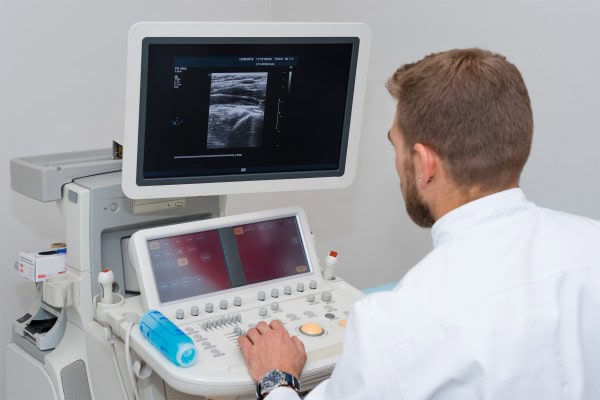
Diagnostic ultrasound has since its clinical introduction in the 1950s, an enormous change. Simple A-Mode devices on the B-Mode, up to highly complex and multi-modal systems with AI support – every step in the development of the diagnostic possibilities has expanded and patient care improved. Doctor Bernhard Scheja outlines the major milestones of this development and explains how technological advances in medical practice have changed.
The history of medical ultrasound technology is an impressive success story – from an experimental procedure to an indispensable diagnostic tool in virtually every medical specialty. Bernhard Scheja explains how the technology have shaped breakthroughs on this path and how the patient care revolution. It is clear that each Generation of new ultrasound devices only improved the quality of the image, but also a fundamentally advanced the understanding of physiological and pathological processes.
Of the Sonar technology, high-performance digital ultrasound
The history of the medical ultra begins to sound with Sonar technology that was originally developed for the U-boat tracking in the Second world war. In the 1950s, were the first A-fashion-devices used in medicine, the one-dimensional Amplitudes generated. Internist Bernhard Scheja explains: “These early devices were only able to interfaces between tissues represent a real image, it was still far away.”
A major breakthrough came with the development of the B-Mode method (Brightness-Mode) in the 1960s, which enabled, for the first time two-dimensional cross-sectional images. The devices were bulky and the picture quality for today’s standards, extremely coarse.
The real-time Revolution of the 1970s
The 1970s and 1980s, with the introduction of real-time sonography another revolutionary step. The first moving pictures were shown in real time, which allowed for the investigation of moving organs such as the heart and the basis of the Doppler-sonography put.
In this period, the first special transducers for various applications have been developed areas. Sector transducers, Linear transducers and Convex transducers allowed for a targeted investigation of different regions of the body.
The digital tipping point
The digitization in the 1990s changed the ultrasonic technology is fundamentally. Dr. Bernhard Scheja emphasized: “The digitalization has not only improves the image quality, but also completely new application possibilities created by the 3D-sonography to computer-aided image analysis.”
The Transition from analogue to digital devices also allowed the storage and post-processing of the images. Ultrasound studies have now documented, archived, and with the previous recordings will be compared.
Clinical milestones in different fields
Parallel to the technical development of ultrasound has set numerous clinical milestones that have changed the way the medical practice.
Revolution in obstetrics
The introduction of the prenatal ultrasound diagnosis in the 1970s revolutionized the world of obstetrics. Malformations were detected at an early stage, which identified risk pregnancies and fetal development to be monitored. This led to a significant reduction in perinatal morbidity and mortality.
Breakthrough in Internal medicine
In the Department of Internal medicine, abdominal sonography allowed for the first time, a non-invasive view of the internal organs. The diagnosis of cysts, tumors, or stone disease has changed radically, and many of the invasive diagnostic procedures could be avoided.
Visualization of blood rivers
The introduction of Doppler sonography in the 1980s, opened a completely new dimension. Dr. med. Bernhard Scheja explains: “With the Doppler, we could not see suddenly anatomical structures, but also the blood flows to measure and visualize. This was a real paradigm shift in the diagnostics of the vessels.“
This technology revolutionized the angiology and cardiology. Most osierungen of vessels, thrombosis, or Shunts could now be diagnosed non-invasively.
Technological breakthroughs and their impact
The Evolution of ultrasound technology has been dominated by a number of technological breakthroughs, each of which has specific effects on patient care were.
High-resolution imaging of superficial structures
The development of high-frequency transducers has enabled the presentation of superficial structures with unprecedented precision. These areas of expertise as dermatology, vascular surgery, rheumatology, where fine anatomical Details are crucial.revolutionized
Contrast agent ultrasound
The introduction of the contrast agent, ultrasound has opened new possibilities in the diagnosis of tumors and perfusion representation. Doctor Bernhard Scheja, explains: “With ultrasound contrast agents, we can recognize today, circulation patterns, which are characteristic for certain types of cancer are radiation exposure and with minimal side effects.”
This technology has particularly revolutionized the liver diagnostics, where different tumor entities can be distinguished on the basis of their characteristic accumulation pattern.
Groundbreaking innovations for patient care
The following technological innovations have improved patient care, particularly sustainable:
- 3D/4D ultrasound: Allows spatial representations and movements in real time
- Elastography: Measures and visualizes the tissue stiffness and enhances the tumor diagnostics
- Fusion imaging: Combined ultrasound images with CT or MRI data for precise interventions
- High-resolution micro-display jar: Visualized the smallest vessels without contrast medium
- AI-based image analysis: Assists in the diagnosis and increases the diagnostic accuracy
Dr. Bernhard Scheja: miniaturization as a pioneer for the Point-of-Care ultrasound
A particularly important development strand of the last years, the increasing miniaturization of ultrasound devices. Specialist Dr. Bernhard Scheja sees this as a crucial paradigm shift: “With the availability of compact, portable devices, it has the sound of a stationary diagnostics in the function area to a widely available Point-of-Care processes.”
Immediate Life-Saving Diagnosis
Life-threatening conditions such as Pericardial tamponades, Pneumothorax, or intra-abdominal bleeding can be detected thanks to the mobile ultrasound devices immediately and treated. The FAST Protocol (Focused Assessment with Sonography for Trauma) is now the Standard in emergency medicine and has improved the care of trauma patients is fundamental.
Global dissemination of sonography
Also in resource-poor regions, the availability of portable, affordable ultrasound machines has improved the medical care is essential. Diagnostic possibilities that were previously reserved for specialized centers, today in rural areas or developing countries.
From the analog Signal to artificial intelligence
The digital Transformation has changed the ultrasound diagnosis in the last few decades – from the analog signal processing on the digital image processing to Integration of artificial intelligence.
Digital Image Enhancement
The digital signal processing allows for a continuous post-processing of the images, for example, by gain settings, smoothing algorithms or Speckle reduction. This leads to a significantly improved image quality and Interpretation also makes it easier to more difficult findings.
Networking and data communication
The storage and exchange of digital images have revolutionized the documentation and communication. Findings can be stored in electronic patient records, shared with colleagues or for follow-up used.
The standardized image archiving PACS systems allows for quick and remote access to the results of the investigation. Bernhard Scheja emphasized that this change has not only expanded the technical possibilities, but also changed the way how Doctors work with the ultrasound.
AI-assisted diagnostic ultrasound
Particularly promising is the Integration of artificial intelligence in ultrasound diagnostics. AI Algorithms can help with image optimization, automatic measurements, or in the detection of pathological changes in support.
Dr. med. Bernhard Scheja sees the combination of high-resolution imaging, digital post-processing, and AI-based analysis of great potential: “Artificial intelligence will not replace the doctor, but it can help us findings to collect fast and accurate not to miss any relevant changes.”